


Neuroscience


Not Knowing
Rhawn Gabriel Joseph, Ph.D.
Gnosis refers to knowledge, to know, and originally referred to the acquisition and seeking of spiritual truths. By contrast, an agnostic, is someone who does not know and who lacks spiritual knowledge. Agnosia, therefore, refers to a loss of knowledge and in ability to know. However, whereas gnosis refers to a spiritual condition, agnosia is a neurological condition which results in an inability to know, to name, to identify, and to extract meaning from visual, auditory, or tactile impressions.
For example, a patient with agnosia might be unable to recognize a "cup" or a "pair of glasses," and when asked to name these items, might mistakenly identify them as a "clock" or "jewelery."
Agnosia, however, is not a naming disorder ("anomia"). Individuals with anomia have difficulty naming an object if it is presented by touch, by sound, via visual inspection, or through auditory description. Individuals may have agnosia limited to a single or multiple modalities. If shown a comb they may be unable to name or describe it's use. However, if the comb were placed in their hand and they were encouraged to palpate and tactually explore it, they may be able to identify it and name it without difficulty. However, if the damage were in the parietal lobe, patients will be able to recognize an item by sight, but not by touch alone. Moreover, if you were to supply the correct name and then show them the items a few minutes later, they will again be unable to identify these objects.
There are a variety of agnosic conditions, which in turn are due to damage to different regions of the cerebrum. For example, patients may suffer an agnosia for environmental, emotional or musical sounds following a right (middle-superior) temporal lobe injury. By contrast, patients may suffer an auditory agnosia for words and sentences following a left superior temporal lobe injury, or a visual agnosia following a damage to the middle-inferior temporal lobe, or inferior-parietal occipital lobe. In some instances, patients may be unable to recognize faces, including the faces of friends, family, or even their own face in the mirror--a condition referred to as "prosopagnosia." In some cases, the agnosia be quite circumscribed and limited to emotion and social signals. These latter disturbances, collectively referred to as a "social-emotional agnosia" may occur with prosopagnosia and are also stereotypically associated with right cerebral injuries.
In some cases, the agnosia is due to a "disconnection syndrome" such that the language areas of the brain become disconnected from the areas of the brain which are perceiving. This is why a patient may be unable to name or recognize an object shown to them visually, but can readily name the same object if it is placed in their hand. The tactile route to the language areas is intact.
In other instances, however, agnosia is due to destruction of the tissue which normally recognizes these stimuli. For example, if the visual feature detectors, which respond to specific shapes and images, are destroyed, those shapes and images can no longer be recognized.
VISUAL AGNOSIA
Visual agnosia is a condition where the patient loses the capacity to visually recognize objects, although visual sensory functioning is largely normal. That is, objects are detected but ability to evoke or assign meaning is lost and the object cannot be identified. The percept becomes stripped of its meaning.
For example, if shown a comb, the patient might have no idea as to what it is or what it might be used for. However, they may be capable of giving a rough estimate of its size and proportions and other features.
In general, this form of visual agnosia is associated with damage involving the medial and deep mesial portion of the left occipital lobe as well as the basal (middle-inferior) temporal lobe. Indeed, the left posterior basal temporal lobe is a convergence zone for visual, auditory, and tactile information, and becomes activated during a variety of language tasks, including reading and object and letter naming (Price, 2007). This has been demonstrated not only by functional imaging, but direct cortical recording, and electrical stimulation.
APPERCEPTIVE & ASSOCIATIVE VISUAL AGNOSIA
"Apperceptive visual agnosia" refers to a disturbance in perceptual and visual-motor integration, such that patients have difficulty copying or matching various objects. This latter form of agnosia has been associated with lesions to the parietal occipital cortex (Mizuno, et al., 1996), as well as to bilateral damage to the inferior-occipital cortex (Shelton, et al., 1994).
By contrast "associative visual agnosia," is due to disconnection from the language area, such that auditory equivalents (or the names of) visual items cannot be matched to a visual perception.This is usually associated with left inferior and middle temporal (area 37) occipital abnormalities, and to lesions to and atrophy of the parietal occipital cortex.
SOCIAL-EMOTIONAL AGNOSIA
Social-emotional agnosia is due to a right cerebral, or bilateral temporal and amygdala injury. Patients typically are unable to correctly perceive or comprehend social-emotional nuances conveyed through voice, gesture, or facial expression.
Patients diagnosed with "schizophrenia" and children afflicted with autism, often appear to suffer from abnormalities involving social-emotional perceptual and expressive functioning, and amygdala and temporal lobe abnormalities have been found in these populations. If due to amygdala and temporal lobe injury, this may explain why these individuals appear to have extreme difficulty determining and identifying the motivational and emotional significance of externally occuring events, to discern social-emotional nuances conveyed by others, or to select what behavior is appropriate given a specific social context. In consequence, in addition to their other afflictions, autistic children or schizophrenic adults may appear socially emotionally blunted or agnosic to varying degrees.
Social-emotional agnosias are commonly observed following amygdala, and amygdala, and right cerebral lesions involving the temporal lobe in particular (Joseph 1988a, 1992a). For example, with damage to the right temporal-occipital region, there can result a severe disturbance in the ability to recognize the faces of friends, loved ones, or pets (Braun et al. 1994; DeRenzi, 1986; DeRenzi et. al., 1968; DeRenzi & Spinnler, 1966; Hanley et al. 1990; Hecaen & Angelergues, 1962; Landis et al., 1986; Levine, 1978; Whitely & Warrington, 1977l ) or to discriminate and identify even facial affect (Braun et al. 1994); prosopagnosia. In fact, with gradual deterioration and degeneration of the right inferior temporal lobe, patients may suffer a progressive prosopagnosia (Evans et al. 2005). Some patients may in fact be unable to recognize their own face in the mirror.
In large part these neocortical deficits are due to limbic system disconnection, and/or destruction of specific limbic nuclei such as the amygdala (Joseph 1992a; see chapter 13). For example, lesions that destroy amygdala fibers of passage to the right or left temporal lobe, essentially disconnect the neocortex and amygdala such that they can no longer receive or transmit appropriate signals. In addition, lesions to only the right or left amygdala (n primates) may induce social-emotional agnosic states, but only in regard to stimuli and persons in the contralateral half of auditory, visual, and tactile space (Downer 1961); e.g. only the right or left hemisphere may become severely effected. Hence, in humans, given right cerebral dominance for social and emotional stimuli, a right sided disconnection or destruction of the amygdala can exert profound influences regardless of which hemisphere is perceiving and responding to emotional stimuli.
Among primates and mammals, bilateral destruction of the amygdala significantly disturbs the ability to determine and identify the motivational and emotional significance of externally occuring events, to discern social-emotional nuances conveyed by others, or to select what behavior is appropriate given a specific social context (Bunnel, 1966; Fuller et al. 1957; Gloor, 1960; Kling & Brothers 1992; Kluver & Bucy, 1939; Weiskrantz, 1956). Bilateral lesions lower responsiveness to aversive and social stimuli, reduce aggressiveness, fearfulness, competitiveness, dominance, and social interest (Rosvold et al. 1954). Indeed, this condition is so pervasive that subjects seem to have tremendous difficulty discerning the meaning or recognizing the significance of even common objects -- a condition sometimes referred to as "psychic blindness", or, the "Kluver-Bucy syndrome".
Thus, animals with bilateral amygdaloid destruction, although able to see and interact with their environment, may respond in an emotionally blunted manner, and seem unable to recognize what they see, feel, and experience. Things seem stripped of meaning (Brown & Schaffer, 1888; Gloor, 1960; Kluver & Bucy, 1939; Weiskrantz, 1956) such that even the ability to appropriately interact with loved ones is impaired; similar, in some respects to what occurs in autism and with adults afflicted with schizophrenia.
For example, Terzian & Ore (2011) described a young man who following bilateral removal of the amygdala subsequently demonstrated an inability to recognize anyone, including close friends, relatives and his mother. He ceased to respond in an emotional manner to his environment and seemed unable to recognize feelings expressed by others. In addition, he became extremely socially unresponsive such that he preferred to sit in isolation, well away from others.
Among primates who have undergone bilateral amygdaloid removal, once they are released from captivity and allowed to return to their social group, a social-emotional agnosia becomes readily apparent as they no longer respond to or seem able to appreciate or understand emotional or social nuances. Indeed, they appear to have little or no interest in social activity and persistently attempt to avoid contact with others (Dicks et al. 1969; Jonason & Enloe, 1971; Kling & Brothers 1992; Jonason et al. 1973). If approached they withdraw, and if followed they flee. Indeed, they behave as if they have no understanding of what is expected of them or what others intend or are attempting to convey, even when the behavior is quite friendly and concerned. Among adults with bilateral lesions, total isolation seems to be preferred.
In addition, they no longer display appropriate social or emotional behaviors, and if kept in captivity will fall in dominance in a group or competitive situation -- even when formerly dominant (Bunnel, 1966; Dicks et al., 1969; Fuller et al., 1957; Jonason & Enloe, 1971; Jonason et al., 1973; Rosvold et al. 1954).
As might be expected, maternal behavior is severly affected. According to Kling (1972), mothers will behave as if their "infant were a strange object be be mouthed, bitten and tossed around as though it were a rubber ball".
Given the many similarities between amygdala destruction and the social-emotional agnosias demonstrated by certain subgroups of those classified as schizophrenic, as well as those diagnosed as autistic, it would thus appear that amygdala/temporal lobe dysfunction is implicated as a major source of psychopathology in these patient populations.
FINGER AGNOSIA
Finger agnosia is not a form of finger blindness , as the name suggests. Nor is it an inability to recognize a finger as a finger. Rather, the difficulty involves naming and differentiating among the fingers of either hand as well as the hands of others (Gerstmann, 1940). This includes pointing to fingers named by the examiner, or moving or indicating a particular finger on one hand when the same finger is stimulated on the opposite hand. In addition, if you touch their finger while eyes are closed, and ask them to touch the same finger they may have difficulty. Often patients who have difficulty identifying fingers by name or simply differentiating between them non-verbally also suffer from receptive language abnormalities (Sanquet, et al. 1971). However, disturbances differentiating between the different fingers can occur independent of language abnormalities or with right parietal injuries (in which case the problem is seen only with the patient's left hand).
Usually, however, finger agnosia is associated with left parietal (as well as temporal-occipital) lobe lesions in which case the agnosia is demonstrable in both hands. It is also part of the constellation of symptoms often referred to as Gerstmanns Syndrome, i.e. agraphia, acalculia, left-right disorientation, finger agnosia (Gerstmann, 1942). Gerstmann's symptom complex is most often associated with lesions in the area of the supramarginal gyrus and superior parietal lobule (Hrbek, 1977; Strub & Geschwind, 1983).
SIMULTANAGNOSIA
Simultanagnosia is an inability to see more than one thing or one aspect of an object at a time, although individual details may be correctly perceived. However, the patient is unable to relate the different details so as to discern what is being viewed. For example, if shown a picture of a man holding an umbrella and a suitcase, they may see the suitcase and the man, and the umbrella, but be unable to relate these items into a meaningfull whole. In fact, by surrounding the object with other objects perceptual recognition may deteriorate even further.
With severe damage the patient may be unable to even recognize individual objects. Indeed, one patient I examined with bilateral posterior parietal-occipital damage (following a bilateral posterior artery stroke) could only identify parts of objects, but not the object itself.
In part, this is due to a breakdown in the ability to perform serial feature-by-feature visual analysis, and is sometimes accompanied by abnormal eye movements. Nevertheless, a variety of anatomical regions, when compromised can give rise to this abnormality. For example, simultanagnosia has been noted to occur with left, right and bilateral superior occipital lobe lesions, or injuries involving the frontal eye fields. More over, I have observed patients with diffuse degenerative disturbances who demonstrate simultangnosic deficiencies.
CATEGORY-SPECIFIC AGNOSIA, AND THE RIGHT & LEFT TEMPORAL LOBE
Depending on the laterality and location of the injury, e.g., right vs left inferior/medial vs superior temporal lobe, patients may display category specific agnosias. For example, a 27-year old man I examined who had sustained a massive right inferior-posterior temporal lobe injury that also involved surgical removal, was able to recognize pictures of tools (he had been a carpenter) but could not recognize or correctly name pictures of animals and he could not correctly remember facial stimuli that he had been shown five minutes earlier and could not differentiate them from faces he had not seen. By contrast, a 43 year old woman who had been a waitress, and had developed a left inferior temporal lobe glioma that required surgical removal (coupled with chemotherapy) was able to recognize and name pictures of animals and could remember different pictures of faces, but had considerable difficulty recognizing and naming common household objects.
In this regard a 47-year old woman I examined who was subsequently found to have a calcium cyst growing from the skull into the right superior temporal lobe, was able to name pictures of animals, tools and household objects. However, she was almost completely unable to recognize and correctly name animal and humans sounds (e.g. a baby crying, a crowd cheering, a lion roaring) which had been briefly presented, but was better able to recognize non-living sounds such as a creaking door, or a hammer hammering--though these abilities were also compromised.
These findings, which require independent confirmation, raises the possibility that the temporal lobes are not only able to distinguish between living and non-living things including tools and faces, but that the right temporal lobe is specialized for perceiving living creatures (and the sounds they make) whereas the left is specialized for perceiving and naming non-living things, such as tools and household objects.
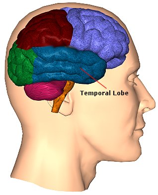
THE TEMPORAL LOBE & THE NEUROLOGY OF FORM RECOGNITION
The neocortex of the inferior temporla lobe is specialized for receiving, analyzing, discriminating, recognizing and recalling complex visual information and is involved in attention and visually guided behavior, including the recollection and learning of visual discriminations (Gross, 1972) and memory for objects and spatial locations (Nunn et al., 2015; Ploner et al., 2015). If the temporal lobe is injured, these functions become compromised.
For example, Kimura (1963) found that patients with right vs left temporal lobe injury were impaired when presented with overlapping nonsense shapes and then immediately tested for recognition. Likewise, Meier and French (1965), found that those with right vs left temporal lobe injuries were impaired when asked to make visual discriminations when presented with fragmented concentric circle patterns--skills which are also related to visual closure and gestalt formation. More recently, Nunn et al., (2015) found that right vs left temporal lobectomy patients were more impaired when required to remember and recognize toys and recall their location.
Cells in the ITL have very large, bilateral visual receptive fields which include the fovea, and many are sensitive to direction of stimulus movement, color, contrast, size, shape, orientation and are involved in the perception of three dimensional objects and the supramodal analysis of information already processed in the association areas. Thus inferior temporal (IT) neurons appear to take part in the last stages of visual analysis for form recognition and receive terminal fibers from the primary and association visual areas; that is, via the ventral visual stream. Indeed, a single neuron can respond to a combination of these features and many will fire selectively in response to particular shapes and faces.
In fact, based on single cell recordings, some of these temporal lobe neurons have been found to become particularly excited when presented with two dimensional patterns or three dimensional objects such as hands, brushes, and in particular, faces (Deismone & Gross, 1979; Gross et al., 1972; Nakamura et al. 1994; Richmond et al., 1983; Richmond et al.2013). A variety of different feature detectors are found and the majority probably act collectively so as code and assemble a particular shape, including the formation of gestalts and thus the performance of visual closure, particularly the right temporal region. Via closure an individual can detect a partial stimulus and determine if the stimulus is that of a human face vs a rabbit. Hence, if these areas are damaged, and these feature detecting neurons are destroyed, the patient will lose the ability to visually recognize a variety of visual stimuli, including the human face.
MUSIC AND NON-VERBAL ENVIRONMENTAL AND ANIMAL SOUNDS
Individuals with extensive left-hemisphere damage and/or severe forms of expressive aphasia, although unable to discourse fluently, may be capable of swearing, singing, praying or making statements of self-pity. Even when the entire left hemisphere has been removed completely, the ability to sing familiar songs or even learn new ones may be preserved (Smith, 1966; Smith & Burklund, 1966) --although in the absence of music the patient would be unable to say the very words that he or she had just sung. The preservation of the ability to sing has, in fact, been utilized to promote linguistic recovery in aphasic patients, i.e., melodic-intonation therapy (Albert et al. 1973; Helm-Estabrooks, 1983).
Similarly, there have been reports that some musicians and composers who were suffering from aphasia and/or significant left hemisphere impairment were able to continue their work (Alajounine, 1948; Critchley, 1953; Luria, 1973). In some cases, despite severe receptive aphasia and/or although the ability to read written language (alexia) was disrupted, the ability to read music or to continue composing was preserved (Gates & Bradshaw, 1977; Luria, 1973).
One famous example is that of Maurice Ravel, who suffered an injury to the left half of his brain in an auto accident. This resulted in ideomotor apraxia, dysgraphia, and moderate disturbances in comprehending speech (i.e., Wernicke's Aphasia). Nevertheless, he had no difficulty recognizing various musical compositions, was able to detect even minor errors when compositions were played, and was able to correct those errors by playing them correctly on the piano (Alajounine, 1948).
Conversely, it has been reported that musicians who are suffering from right hemisphere damage (e.g., right temporal-parietal stroke) have major difficulties recognizing familiar melodies and suffer from expressive instrumental amusia (Luria, 1973; McFarland & Fortin, 1982). Even among nonmusicians, right hemisphere damage (e.g. right temporal lobectomy) disrupts time sense, rhythm, and the ability to perceive, recognize or recall tones, loudness, timbre, and melody . In fact, right temporal injuries can disrupt the ability to remember musical tunes or to create musical imagery (Zatorre & Halpen, 2003).
Right hemisphere damage also can disrupt the ability to sing or carry a tune and can cause toneless, monotonous speech, as well as abolish the capacity to obtain pleasure while listening to music (Reese. 1948; Ross, 1981; Shapiro & Danly, 2015), i.e., a condition also referred to as amusia. For example, Freeman and Williams (1953) report that removal of the right amygdala in one patient resulted in a great change in the pitch and timbre of speech and that the ability to sing also was severely affected. Similarly, when the right hemisphere is anesthetized the melodic aspects of speech and singing become significantly impaired (Gordon & Bogen, 1974).
It also has been demonstrated consistently in normals (such as in dicotic listening studies) and with brain-injured individuals that the right hemisphere predominates in the perception (and/or expression) of timbre, chords, tone, pitch, loudness, melody, meter, tempo, and intensity--the major components (in conjunction with harmony) of a musical stimulus.
For example, in a functional imaging study, it was found that when professional pianists played Bach (Bach's Italian concerto, third movement) that there was increased activity in the right but not left temporal lobe, whereas when they played scales, activity increased in the left but not right temporal lobe (Parsons & Fox, 2007). Likewise, Evers and colleagues (2015) in evaluating cerebral blood velocity, found that a right hemisphere increase in blood flow when listening to harmony (but not rhythm), among non-musicians in general, and especially among females.
In addition, Penfield and Perot (1963) report that musical hallucinations most frequently result from electrical stimulation of the right superior and lateral surface of the temporal lobe. Berrios (1990) also concluded from a review of lesions studies that musical hallucinations were far more likely following right cerebral dysfunction; whereas conversely destruction of this tissue disrupt the ability to conjure up musical imagery (Zatorre & Halpen, 2003). Findings such as these have added greatly to the conviction that the right cerebral hemisphere is dominant in regard to the non-temporal sequential aspects of musical perception and expression.
However, this does not appear to be the case with professional musicians who in some respects tend to treat music as a mathematical language that is subject to rhythmic analysis. As noted when professional pianists played scaled, activity increased in the left but not right temporal lobe (Parsons & Fox 2007). Moreover, Evers and colleagues (2015) found that musicians displayed an increase in left hemisphere blood flow when listening to both harmony and rhythm.
ENVIRONMENTAL AND HUMAN/ANIMAL SOUNDS
In addition to music the right hemisphere has been shown to be superior to the left in discerning and recognizing nonverbal and environmental sounds (Curry, 1967; Joseph, 1988b; Kimura, 1963; King & Kimura, 1972; Knox & Kimura, 1970; Nielsen, 1946; Piazza, 1980; Roland et al. 1981; Schnider et al. 1994; Spreen et al. 1965; Tsunoda, 1975). Similarly, damage that involves select areas within the right hemisphere not only disturb the capacity to discern musical and social-emotional vocal nuances, but may disrupt the ability to perceive, recognize, or disciminate between a diverse number of sounds which occur naturally within the environment (Fujii et al., 1990; Joseph, 2003; Nielsen, 1946; Schnider et al. 1994; Spreen et al. 1965), such as water splashing, a door banging, applause, or even a typewriter; this is a condition which also plagues the disconnected left hemisphere (Joseph, 1988b).
A 47-year old woman I examined who was subsequently found to have a calcium cyst growing from the skull into the right superior temporal lobe, was able to name pictures of animals, tools and household objects. However, she was almost completely unable to recognize and correctly name animal and humans sounds (e.g. a baby crying, a crowd cheering, a lion roaring) which had been briefly presented, but was better able to recognize non-living sounds such as a creaking door, or a hammer hammering--though these abilities were also compromised. However, as detailed above, there is some possiblilty that the right temporal lobe is better able to recognize living and true environmental sounds, whereas the left may be better able to recognize and name non-living sounds, such as those produced by machinery.


DVD - Brain Lectures
Six Lectures
3 DVD set = $29.95(+ shipping)
Pay By Paypal.com
DVD 1: Brain Overview
DVD 2: The Left Hemisphere, Brainstem, Midbrain, Thalamus
DVD 3: The Frontal Lobes: Frontal Lobotomy, Memory, Aphasia, Paralysis
DVD 4: The Parietal Lobes: Touch, Body-in-Space, Body Image, Hemi-Neglect, Phantom Limbs,
DVD 5: The Temporal Lobes: Language, Memory, Aphasia, Hallucinations, Face Recognition
DVD 6: The Limbic System: Amygdala, Hippocampus, Hypothalamus, Sex, Emotion, Memory, Stress, PTSD, Hallucinations