


Rhawn Joseph, Ph.D.
BrainMind.com
For the first several years of life, the nervous system of the infant and child is exceedingly immature and requires considerable social, emotional, physical, and maternal contact in order to develop normally (Joseph, 1982, 1996, 1999ab, 2000). If deprived of this contact, or if the child suffers from periodic neglect or loss of maternal contact, the nervous system will be profoundly affected and all aspects of social, emotional and psychological functioning will be profoundly effected (Ainsworth, et al., 1978; Bowlby, 1982; Cicchetti & Tucker, 2004; Joseph, 1982, 1996, 1999ab; Koluchova, 2006; Langmeier & Matejcek, 1975; Lanting et al., 2015; Spitz, 1945). Even the infant's immune system can be injured and the child may become ill, and may die. As noted, Angela also became ill with some "diseases" and she was apparently so ill that several parental visits were cancelled. Moreover, many of Angela's fantasies center on death.
Because the infant requires loving maternal input, the greater the degree of maternal deprivation, the more serious might be the consequences. For example, in a review of the world wide literature, the mortality rates for infants placed in foundling homes was found to be over 70% (Langmeier & Matejcek, 1975)--a consequence not of poor nutrition, but lack of maternal care from the biological mother. Likewise, it has been found that the risk of infant motorality increases by a factor of five if the mother dies when the infant is between 6 months to one year of age, even if care is provided by female relatives (Hill & Hurtado 1996). In part, this is due to the effects of stress induced by maternal separation, and the stress-release or cortisol which can attack the immune system resulting in illness and death (Joseph, 1999ab).
THE IMMATURE BRAIN REQUIRES MATERNAL CONTACT
Because of the tremendous need for maternal stimulation, even subtle differences in the degree and quality of maternal contact can differentially effect the brain and cognition and can cause neurological abnormalities and cognitive-motor disturbances (Lanting et al., 2015; Joseph, 2003). As noted, Angela displays evidence of right cerebral dysfunction, reduced non-verbal intellectual ability, attention deficits, hyperactivity, and non-verbal memory deficits.
As noted, Angela's contact with her mother was severely restricted, and she was allowed contact with her mother inconsistently, approximately once per month. As will be detailed, inconsistent maternal contact, and prolonged periods of separation between the ages of 1-3, can exert profound effects on social, emotional, and even sexual functioning (Ainsworth, et al., 1978; Bowlby, 1982; Cicchetti & Tucker, 2004; Joseph, 1982, 1996, 1999ab; Koluchova, 2006; Langmeier & Matejcek, 1975; Lanting et al., 2015; Spitz, 1945); and these deficits, in turn are a result of injury to the brain secondary to insufficient stimulation and the damaging effects of stress on the brain Joseph, 1998, 1999ab; Lupien & McEwen, 1997; Sapolsky, 1996). As noted, the brain is exceedingly immature. Connections between neurons, and the growth of the neurons and their axons and dendrites are directly affected by the amount and quality of the stimulation received. The brain of the infant, including a child between the ages of 1-3, requires considerable age-appropriate, "experience-expectant" social emotional, maternal, and perceptual stimulation to develop normally, including considerable face-to-face and eye-to-eye contact from the primary caretakers, i.e. the mother. If sufficient normal stimulation is not provided, or deprived of consistent maternal contact, developing neurons and dendrites will establish abnormal interconnections, or they will whither, die, and drop out at an accelerated rate (Dimond, 1991; Greenough & Black, 1993; Joseph, 1999b; Siegel et al., 1993). This is called the "use it or lose it principal." If insufficiently stimulated, or if deprived of or provided insufficient maternal care, brain cells begin to die or they may develop aberrant interconnections. Angela was denied normal maternal contact for 7 months.
Different parts of the developing brain, however, begin to mature at different times. For example, the right hemisphere is more vulnerable to long term damage, beginning after the first year whereas the left hemisphere is more vulnerable during the first year (Novelly & Joseph, 1983; Joseph, 1982, 1986). However, the limbic system remains highly vulnerable for the first several years. Likewise, because the developing brain is "experience-expectant," these brain areas require specific types of maternal and other forms of stimulation at specific times or damage and injury will result.
Limbic system structures mediate all aspects of social and emotional functioning (Joseph, 1982, 1990, 1992, 2004, 1996, 1998, 1999ab). As the limbic system matures, these structures require "experience expectant" maternal and emotional stimulation in order to develop normally (Joseph, 1982, 1996, 1998, 1999ab). If that stimulation is not provided, the infant may experience incredible stress, and emotional and stress-induced abn�rmalities result (Joseph, 1998, 1999ab; Kraemer, 1992; Lupien & McEwen, 1997; Sapolsky, 1996; Diamond, 1985, 1991; Kraemer, 1992; Siegel, et al., 1993; Walsh, Budtz-Olsen, Penny, & Cummins, 1969).
The neocortex, which mediates all aspects of higher cognitive functioning, including verbal and non-verbal intelligence, also require "experience expectant" stimulation in order to develop normally (Greenough & Black, 1992). If that stimulation is not provided, abn�rmalities result.
Indeed, it is well established that early environmental influences exert significant organizing effects on neural growth throughout the neocortex and limbic system, determining neocortical thickness, the density, size, shape and growth of dendrites and synapses, and the die off and drop out rate of glia, neurons, and axons (Diamond, 1991; Greer, Diamond, & Tang, 1982; Greenough & Black, 1992; Siegel et al., 1993).
Initially synapses and interconnections may be formed randomly and in excess numbers, such that those which are not stimulated drop out. That is, synapses and neural interconnections are established at a certain time period in expectation that experience will activate them at the appropriate time. As summed up by Greenough et al. (1987, p. 540) "since the normal environment reliably provides all species members with certain experiences, many mammalian species have evolved neural mechanism that take advantage of such experiences to shape developing sensory and motor systems. An important component... appears to be the intrinsically governed generation of an excess of synaptic connections among neurons, with experiential input subsequently determining which of them survive."
If deprived of these normal early experience during specific critical developmental periods this can result in the elimination of millions of neurons, axons, dendrites and synapses, and may even induce functional invasion by competing neuronal cell assemblies which establish functional dominance over those neurons that have been insufficiently or adversely stimulated (Casagrande & Joseph, 1978, 1980; Hirsch & Spinelli, 1971; Joseph, 1986b; Spinelli et al., 1972).
As demonstrated in animal (Geyer et al., 1993; Hirsch & Spinelli,1971; Spinelli et al., 1972) and in human studies (Joseph, 1986b; Novelly & Joseph, 1983), neocortical and limbic neurons deprived of normal experience sometimes come to subserve wholly different functions. In consequence, even when normal experience is later provided, deprived neurons are unable to function normally.
Children (Ainsworth, et al., 1978; Bowlby, 1982; Cicchetti & Tucker, 2004; Koluchova, 2006; Spitz, 1945) and non-human primates and mammals (Geyer et al., 1993; Harlow & Harlow, 1965a,b; Joseph, 1979; Joseph & Casagrande, 1980; Joseph & Gallagher, 1980) reared under maternally deprived, or neglectful conditions, suffer severe social, emotional, cognitive, perceptual, and intellectual deficits. These disturbances include a diminished ability to anticipate consequences or to inhibit irrelevant or inappropriate behaviors, impulsiveness, hyperactivity (Joseph & Gallagher, 1982), and an impaired ability to form loving attachments (Ainsworth, et al., 1978; Bowlby, 1982; Cicchetti & Tucker, 2004; Koluchova, 2006; Spitz, 1945).
Likewise, in the present case, Angela demonstrates hyperactive behaviors, impulsiveness, anxiety, and disturbances in her ability to feel emotionally attached.
Even the ability to function in a sexually normal fashion is disrupted (Fritz & Fritz,1985; King & Mellen, 2004; Langmeier & Matejcek, 1975; Moberg, 1985). Sexuality often becomes abnormal, because sexuality is associated with the functional integrity of the limbic system (the amygdala, septal nuclei, and hypothalamus in particular). However, deficits in sexuality may not become manifest until adulthood or the teenage years.
THE NEED FOR MOTHERING & MATERNAL CONTACT COMFORT
Because of the immaturity of the brain and the incredible need for social, emotional contact, for the first 8 months of life children show no fear of strangers and generally respond by smiling and with pleasure and happiness when strangers approach and pick them up (Charlesworth & Kruetzer, 1973; Spitz & Wolf 1946; Sroufe, 1996). For the first 8 months, the child shows indiscriminate friendliness which increases the likelihood that adults and especially the mother, will provide the stimulation and contact comfort which the nervous requires in order to develop normally (Joseph, 1982, 1996, 1998, 1999ab). However, during the first 8 months, the infant also develop a bond to its mother and becomes increasingly attached to the mother.
Nevertheless, so strong is the need for contact comfort and maternal contact, that young animals raised in social isolation will form attachments to bare wire frames, inanimate objects, and television sets, and will seek out animals that might maul them and creatures that might eat them (Cairn, 1966; Fisher, 1955; Harlow & Harlow 1965a,b; Hoffman & DePaulo, 1977). So powerful is the need for contact comfort, particularly with the mother, that infant humans, non-human primates, and a variety of young creatures will desperately seek contact with mothers and caretakers who reject, physically abuse, and who nearly kill them.


The immature "experience- expectant" brain in fact demands emotional and social contact, especially from the mother, and will seek it out regardless of consequences. Even an abusive mother is better than a mother substitute (Bass et al., 2004; Harden, 2004; Kortenkamp, K., and Ehrle, 2002; Lawrence et al. 2006 NSCAW 2004).
In fact, studies show that even abused children placed in foster care do more poorly than abused children who remain with their abusive parents (Bass et al., 2004; Harden, 2004; Kortenkamp, K., and Ehrle, 2002; Lawrence et al. 2006 NSCAW 2004).
EFFECTS OF MATERNAL SEPARATION ON SOCIAL-EMOTIONAL FUNCTIONING
Children between the ages of 1 and 3 who are reared in institutions where mothering and emotional comfort are minimized typically become incredibly upset, will scream and cry, develop emotional abnormalities, and later show signs of intellectual deficits, severe attentional deficits, disturbed ability to form emotional attachments, and exceedingly abnormal social behavior (Dennis, 1975; Goldfarb 1943, 1945, 1946; Langmeier & Matejcek, 1975; Spitz, 1945, 1946). Likewise, Angela was denied normal maternal contact during this time period, for 7 months, and she too would cry when separated from her mother, and she too developed emotional disturbances, reduced non-verbal compared to her verbal intellectual ability, hyperactivity, attentional deficits, and a disturbed ability to form emotional attachments.
R.A. Spitz (1945, 1946) found that deterioration is in fact quite rapid among children removed from their mothers between the ages of 1 and 3. As the separation became prolonged, unmothered children would frequently cry but became unresponsive to social stimulation, would lay passively on their beds, and made vigorous attempts to avoid strangers or novel objects or toys. When approached they would scream and withdraw. These deprived youngsters spent hours engaged in obsessive, repetitive, stereotyped, and abusive self-stimulating movements; i.e. head banging, rocking, or repeatedly pinching the same piece of skin until sores developed.
Nor are these catastrophic consequence limited to humans, for similar disturbances characterize the behavior of other primates reared under deprived maternal conditions. As detailed by Harlow and Harlow (1965; p. 138), monkeys raised alone, in a sterile environment, with surrogate terry cloth mothers would stereotypically "sit in their cages and stare fixedly into space, circle their cages in a repetitive stereotyped manner and clasp their heads in their hands or arms and rock for long periods of time. They develop compulsive habits, such as pinching precisely the same patch of skin between the same fingers hundreds of times a day; occasionally such behavior may become punitive and the animal may chew and tear at its body until it bleeds."


Children removed at this critical age, and who received inconsistent, limited, and restricted maternal contact, as in the present case, also develop emotional, intellectual, and neurological abnormalities.
Children between the ages of 9 months to 3 years, who are removed from their mothers, even for brief periods, suffer extreme separation anxiety, and they experience incredible stress as demonstrated by their crying when separated. As will be detailed later, stress can damage the brain and the immune system, via the secretion of cortisol.
Studies on infants where "mothering" is inconsistent, as in the present case, have found that the child may feel awkward and uncomfortable when around others, and may be easily frightened, easily stressed, and may forever fear abandonment. Moreover, the child's ability to form healthy emotional attachment, during childhood or adulthood, may become abnormal and their ability to relate successfully with others will become disturbed (Ainsworth et al., 1978; Bowlby, 1960, 1982; Cicchetti & Tucker, 2004; De Wolff & Ijzendoorn, 1997; Isabella, 1993; Izard & Harris, 2015; Kerns & Barth, 2015; Sroufe, 1996). Thus, the prognosis, in the present case, is most negative.
MATERNAL BONDING & STRANGER FEAR
Between the ages of 9-12 months, infants increasingly become upset at the prospect of maternal separation (Ainsworth et al., 1978; Bowlby 1969, 1982; Schaffer, 1966; Spitz, 1946). They also begin to experience and vocalize fear and separation anxiety (Bronson, 1972; Emde, Gaensbauer, & Harmon, 2006; Sroufe & Waters, 2006).
By nine months of age, 70% of children respond aversively if a stranger approaches and might cry out (Bronson 1974; Schaffer, 1966; Waters et al., 1975). By 1 year 90% of children respond aversively to strangers (Schaffer, 1966).
In fact studies of children who have been deprived of maternal contact between the ages of 1 and 3, often suffer profound emotional injury which can last a lifetime (Goldfarb, 1945,1946; Koluchova,2006; Spitz, 1946). Depending on the length of the separation, they may display an insatiable need for social stimulation, but also behave in a socially bizarre, bullying, sadistic, and explosively violent fashion. In part this is because of the need for maternal contact, coupled with fear of strangers and the abnormalities which result when deprived of that contact and placed with strangers for prolonged time periods.
Likewise, in the present case, Angela seeks social stimulation, but has also displayed bursts of anger, and engaged in threatening behavior. Angela has exceedingly violent thoughts and her fantasies involve death and destruction.
It has been found that children placed in foundling homes for long time periods after they have reached 9 months of age may respond to strangers with fear which then alternates with extreme social stickiness. The child may withdraw in fear, but then express an intense desire for social contact. They may hug, cuddle, and kiss indiscriminately, display an insatiable need for attention and affection, as well as behave in a fearful, shy, unfriendly, inappropriate and socially bizarre fashion, or they may alternate between fearfulness or aggressiveness and may withdraw when approached or react in an explosively violent fashion (Goldfarb 1943, 1945, 1946; Langmeier & Matejcek, 1975; Spitz 1945; Koluchova, 2006 Langmeier & Matejcek, 1975).
Later in life, even after returned to the mother, the child may become pathologically shy, or they may respond to strangers with extreme stickiness and persistently express an intense desire for social cohesion, which in many cases may include bullying, and abnormal or inappropriate sexual behavior, including unsolicited hugging and kissing, touching and sucking on genitals, or exposing one's self and even urinating on other children (Goldfarb, 1945,1946; Koluchova,2006; Spitz, 1946). That is, they crave social stimulation while simultaneously withdrawing from social stimulation and/or behave in a socially, emotionally, and sexually inappropriate and sexually aggressive fashion.
INCONSISTENT MATERNAL CONTACT IS PROFOUNDLY STRESSFUL:
THE EFFECTS OF STRESS ON THE BRAIN
Maternal deprivation and neglect and inconsistent mothering is exceedingly stressful (Bowlby, 1960, 1982; Pankseep, Normansell, & Herman, 1988; Kraemer, 1992; Rosenblum, Coplan, & Friedman, 2004; Rots, et al., 2015; Suchecki, et al., 1993; Spitz, 1945, 1946). Prolonged stress will damage the brain, and the immature brain is particularly subject to the damaging effects of stress (Joseph, 1998, 1999ab; 2003).
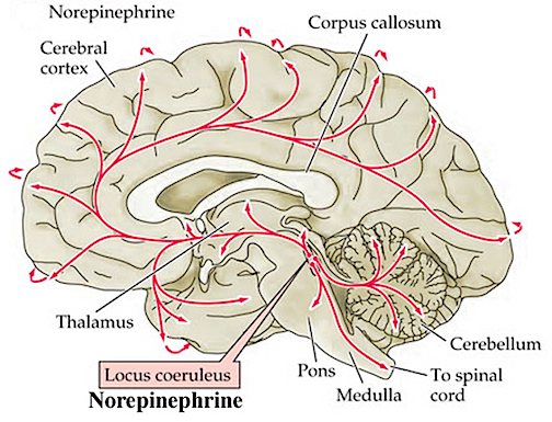
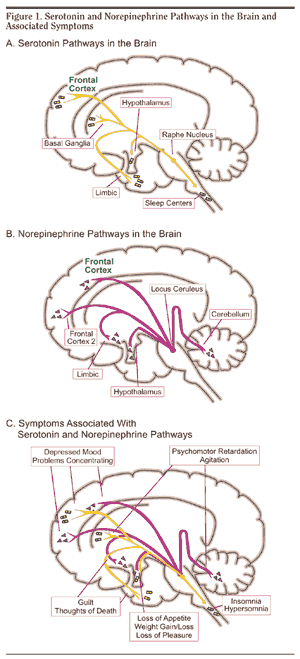
Even temporary periods of maternal separation may trigger significant fluctuations and reductions in neurotransmitters such as norepinephrine (NE), serotonin (5HT) and dopamine (DA), and promote the secretion of enkephalins (Opiate-like substances) and corticosteroids, i.e. cortisol (Kehoe, Clash, Skipsey, & Shoemaker, 1996; Kraemer 1992; Pankseep et al., 1988; Rosenblum et al., 2004).
Reductions in NE and 5HT are associated with the development of emotional abnormalities and severe depression. Alterations in DA are linked to the development of schizophrenia and Parkinson's disease. Excessive secretions of enkephalins are linked to the development of addictive-personalities. Excessive secretions of cortisol can damage the brain, alter the hypothalamus and stress response such that even mild stresses are experienced as extremely stressful, and contributes to the development of Post Traumatic Stress Disorder, PTSD (Joseph, 1996, 1998, 1999ab).
Repeated and prolonged instances of stress will reduced NE and damage the brain. Reductions in NE can severely damage the developing brain because NE plays an exceedingly important role in maximizing neural growth and neuroplasticity (Kasamatsu & Pettigrew 2006; Pettigrew & Kasamatsu 1978; Johnston 1988; Parnavelas et al. 1988). Angela was subjected to repeated and prolonged episodes of stress secondary to maternal separation and as witnessed by her crying when the mother was repeatedly forced to leave after a short visit.
In the developing nervous system, NE also acts to suppress the establishment of irrelevant neural networks and pathways, while simultaneously stabilizing and/or promoting the growth and formation of relevant synaptic circuits (Bear & Singer 1986; Pettigrew & Kasamatsu 2018).
Similarly, NE has been shown to inhibit or suppress irrelevant background activity, while simultaneously enhancing evoked responses in both inhibitory and excitatory circuits associated with the processing of relevant environmental input (Foote et al. 2013). If abnormal, the child may become hyperactive or easily distracted.
The NE neurotransmitter system is also directly implicated in modulating the stress response, and in fact stress increases NE turnover in the amygdala (Tanaka, et al. 2012), a limbic system structure which mediates the fear response, the desire for emotional contact, and which becomes highly active under conditions of emotional and physical stress (Henke, 1992; Ray, et al. 2017; Roozendall, 2014). In fact, the release of NE serves a neural protective function within the amygdala (Glavin, 1985; Ray et al., 1987b). Thus reductions in NE may induce damage to this structure, or the development of abnormal kindling activity under conditions of stress and high arousal.
Unfortunately, NE depletion and excessive amygdala activation such as due to stress, separation anxiety, and stranger fear, can lead to permanent structural and functional alterations within infant and adult amygdala neurons, effecting their neocortical interconnections, postsynaptic densities and in the size of the presynaptic terminals as well as their capacity to process and transmit information (e.g. Cain 1992; Racine 1978).
Because the amygdala mediates all aspects of social emotional functioning, including sexuality and the ability to form normal emotional bonds, injury to this structure will cause profound social-emotional abnormalities such as is characteristic of children who received insufficient mothering and maternal contact during the first 3 years of life (Joseph, 1996, 1998, 1999ab).
In fact, NE reductions result even under mild conditions of neglect (Higley et al. 2012), and even following brief periods of maternal separation (Kraemer et al. 1989) as was the case with Angela Smith. NE appears is directly involved in the experience of separation anxiety from the mother (Pankseep et al. 1988).
It has been shown that if a primate mother is periodically prevented from attending to her infant, the infant will suffer significant reductions in NE activity (Kraemer et al. 1989). Moreover, these infants in turn become less securely attached, more easily frightened and startled, are less social or independent and display significant NE-related abnormalities. According to Rosenblum et al. (2004) infants reared under these mildly stressful conditions display responses similar to those seen in humans suffering from post traumatic stress disorder (PTSD). Thus, infants subjected to the same conditions as Angela Smith, can in fact develop PTSD.
Under stressful conditions not only limbic system structures such as the amygdala and hypothalamus, but the hippocampus may be injured (Joseph, 1996, 1999ab), the hippocampus of the right hemisphere in particular (Joseph, 2003). As the hippocampus acts to place short-term memories into long-term memory (Milner, 1970), stress-induced damage to this structure can effect learning and memory, non-verbal memory in particular (Joseph, 2003). Likewise, stress experienced during early childhood will more greatly affect the right hemisphere and related non-verbal intellectual abilities, such that the PIQ will be significantly lower than the VIQ (Joseph, 2003). These published research findings mirror the deficits demonstrated by Angela Smith.
STRESS DAMAGES THE BRAIN & MIND: CORTISOL, STRESS & EMOTION
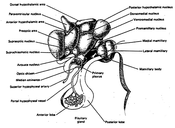
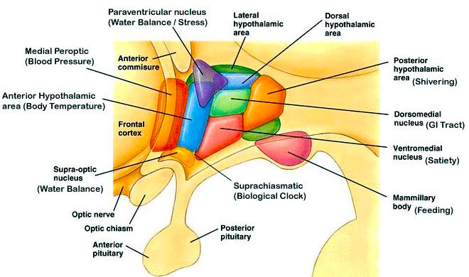
In response to fear, anger, anxiety, neglect, maternal separation, and stress, the anterior hypothalamus begins to secrete corticotropin releasing factor
(CRF) which activates the andenohypophysis which begins secreting ACTH which stimulates the adrenal cortex which secretes glucocorticoids and mineralalocorticoids. The primary glucocorticoid is cortisol (which mobilizes free fatty acids from adipose tissue, breaks down proteins, and increases blood pressure) and the primary mineralocorticoid is aldosterone (Fink, 1999; Hakan, Eyle, & Henriksen, 2004; Moller et al., 1998; Roozendall, Koolhaas & Bohus, 1992). These events are under the modulating influences of the amygdala amd locus coerluesus system which produces norepinephrine (NE).
As stress increases, NE levels are altered and initially exaggerated (Rosenblum et al., 2004; Southwick et al., 1993) only to decrease (Bliss, Ailion, & Zwanziger, 1968; Spoont,1992; Witvliet 1997), which triggers the hyperactivation of the HPA axis and thus the hyper secretion of corticostereoids (Fink, 1999; Moller et al., 1998). With repeated instances of stress, such as due to fear, neglect or separation anxiety, the HPA axis may become hypersensitive and hyperreactive, which in turn can injure the brain, the immune system, and can lead to PTSD.
Since NE also serves a neural protective function (Glavin, 1985; Ray et al., 1987b), if NE levels are reduced--such as due to chronic or repeated instances of severe stress--the continuing hypersecretion of corticosteroids can severely injure pyramidal cells throughout the brain, including the neocortex and limbic system (Gahwiler 1983; Henriksen et al., 1978; Packan & Sapolsky, 1990), including the HPA axis (Rots et al., 2015; Suchecki, et . 1993). Stress and cortisol can also damage neurons located in the hypothalamus, amygdala, and hippocampus (Joseph, 1996, 1998b, 1999b,d; Lupien & McEwen, 1997; Sapolsky, 1996; Uno, Tarara, Else, & Sapolsky, 1989)
If the hypothalamus and the HPA axis is injured, the result may be chronic depression, or sadness, and a host of related emotional abnormalities (see Carrol et al., 2006; Sachar et al. 1973; Swann et al. 2004), including a tendency to forever hyper secrete glucocosteroids and to maintain high levels of cortisol even under neutral conditions. Thus, those effected can subsequently become easily stressed and emotionally upset, anxious, or fearful, even under non-stressful conditions.
Stereotypically, individuals with high cortisol levels tend to feel sad and to become easily depressed, as well as easily overwhelmed by even minimal stress. If sufficiently stressed, they may become ill, and they may also die (Uno et al. 1989).
Stress, including the stress of maternal separation, alters NE, and cortisol levels and can result in the permanent elevation of cortisol even under non-stressful conditions.
Primates who have elevated cortisol levels even under non-stressed conditions, are observed to be passive, low status, submissive, and exhibit an "uptight" rigidity in temparment (Johnson et al., 1996). Moreover, primates with elevated cortisol have difficulty coping with even mild stressors, and tend to easily become infected with diseases, and may "waste away and die with no apparent pathological cause" (Johnson et al., 1996, p. 333).
Likewise, children with elevated levels of cortisol even under non-stress conditions, have been found to be socially abnormal, anxious, and fearful (Kagan, et al 1988). These children are less likely to react in an adaptive manner when stressed and are the most likely to freeze-up, to become afraid, and to passively surrender to their fate (Johnson et al., 1996; Kagan et al., 1988).
As emphasized in this report, children and non-human primates who experience repeated instances of maternal separation not only develop a host of emotional abnormalities, but their immune system is effected and they may become sick and may even die. This is due to the effects of stress and cortisol on the immune system.
For example, cortisol can decrease the production of lymphocytes by the lymph nodes and thymus gland. Since a primary role of lymphocytes is immunological (e.g. to destroy invading bacteria), excessive cortical secretion can disrupt the immune response thus increasing the likelihood that one may get sick.
Moreover, in addition to changes in emotionality and the ability to cope with stress, even sexuality may become abnormal due to the hyper secretion of corticosteroids and chronic stress (Moberg, 1985; Meyer-Bahlburg, 1993; Reinisch & Sanders, 2012).
As noted, stress and the release of cortisol can directly injure the limbic system and other regions of the brain. Neural atrophy and/or abnormal activity is induced because corticosteroids at high levels can exert a suppressive influences on membrane receptor proteins, thereby altering excitability and information transmission between neurons (Hua & Chen, 1989; Majewska, 2016), and can detach the cellular receptor from its attached protein (Beaulieu, 1987); a condition which interferes with messenger RNA protein transcription and thus the genetics of memory. Incoming messages cannot be acted on, learning cannot take place, and injured or damage cells cannot be repaired due to RNA/DNA interference. In consequence, neurons begin to atrophy.
NEGATIVE EFFECTS OF FOSTER CARE ON EMOTIONAL, INTELLECTUAL & PSYCHOLOGICAL DEVELOPMENT
Despite the protests of her father, and although there was no evidence of paternal or maternal abuse or neglect, Angela was placed among strangers, i.e. Foster Care, for a period of at least 7 months, during a critical stage of social and emotional development and neurological immaturity.
It is now well established that Foster Care can be profoundly injurious to a child's mental health (Harden, 2004; Pediatrics 2000), and that "children in foster care have more compromised developmental outcomes than children who do not experience placement in foster care (Harden, 2004).
In fact, children in foster care are more likely to develop behavioral, educational, and emotional problems than children who are raised by abusive and high-risk parents (Bass et al., 2004; Harden, 2004; Kortenkamp, K., and Ehrle, 2002; Lawrence et al. 2006 NSCAW 2004). In Angela's case, there was no evidence of abuse in the parental home.
Children in foster care are also more likely to suffer from health problems, even though, ostensibly they have better access to health care (Bass et al., 2004; Horowitz, 2000).
Children between the ages of 1 month to 3 years are particularly susceptible to emotional harm from foster care, as they are the most fragile (Bass et al., 2004; Perry, 2002).
Foster care can injure a child's emotional development and can lead to negative development outcomes due to inconsistent nurturing and maternal contact (Cassidy et al. 1996). When placed in Foster Care, and if maternal care or care provided by the primary caretaker is inconsistent or inadequate, such as in the case of Angela, children become more insecurely attached and develop abnormal attachments (Cassidy & Berlin 2014; Zeanah et al. 2011).
As detailed by Harden (2004) children placed in Foster Care and who received inconsistent nurturing care from the primary caretaker, and who "have disrupted attachments to their caregivers, display overly vigilant or overly compliant behaviors, show indiscriminate connection to every adult, or do not demonstrate attachment behaviors to any adult. Children with insecure, �disordered� or �disorganized� attachments may also have many other adverse outcomes that persist throughout childhood, such as poor peer relationships, behavioral problems, or other mental health difficulties." Similar findings have been presented by other scientists (Carlson 1998, Lyons-Ruth, 2016).
Children placed in Foster Homes also exhibit "elevated levels of cortisol compared to children reared by their biological parents" (Harden, 2004). Cortisol is secreted in response to stress, and cortisol not only effects the brain but can injure the immune system.
As detailed earlier in this report, children who are removed from their mothers also have a tendency to become ill, and even to die. Likewise, according to Harden (2004) "Many studies have pointed to the deleterious impact of foster care on children's physical health, cognitive and academic functioning, and social-emotional wellbeing. In the area of physical health, pediatric and public health scholars have documented that foster children have a higher level of morbidity throughout childhood than do children not involved in the foster care system."
Likewise, according to the NSCAW, a significant proportion of children placed in Foster Care, suffer from compromised cognitive, intellectual, and academic functioning (National survey of child and adolescent well-being NSCAW, 2001). Specifically, the NSCAW found that over 1/3 of infants and toddlers and 1/2 of pre-school age children scored in the delayed range of development. Similar findings have been presented by other scientists (Konekamp & Ehrle, 2002, Pears & Fisher, 2005; Swayer & Dubowitz, 2004).
Children placed in Foster Care have higher rates of depression, abnormal social skills, are more impulsive, become more aggressive, and are more insecurely attached and have insecure and disordered attachment behaviors (Clausen et al., 1998; Stein et al., 1996).
Children placed in Foster Care do more poorly than children who are left in high-risk homes where parents are neglectful or abusive (Lawrence et al. 2006) Likewise, behavior problems are worse as compared to those raised by maltreating care givers(Lawrence et al. 2006). Likewise, according to a NSCAW (2003) study, children placed in Foster Care show more compromised social-emotional functioning than children raised in a high risk environment. However, in this case, Angela was not being abused or neglected by her parents. In this regard, and given these findings and the research reviewed above it is thus clear that Angela developed these problems precisely because was placed in foster care.
In fact, in a study released on April 6, 2005, by the Casey Family Programs, Harvard Medical School, the State of Washington Office of Children�s Administration Research, and the State of Oregon Department of Human Services,it was reported that children raised in Foster Homes are highly likely to develop PTSD and that the rates of PTSD among adults who were formerly placed in foster care was twice as high as among U.S. war veterans.
As detailed in the Casey Family study, in addition to the PTSD, 54.4% of adults formerly in foster care were found to be suffering from depression, social phobia, panic syndrome, and anxiety. Moreover, it was found that 80% of adults who had been placed in Foster Care as children, were doing poorly, with a quarter to one third becoming homeless or living below the poverty level.
ABUSE WHILE IN FOSTER CARE:
In states from California to New York, and in counties and cities from Santa Clara County and San Jose, to New York City and Maryland, it has been reported that over 20% of children placed in Foster Care are sexually and physically abused or suffer from profound neglect while in Foster Care (United States District Court, D. Maryland, L.J. By and Through Darr v. Massinga, decision, Civ. No. JH-84-4409, September 27, 1988. F.Supp. 508 (D.Md. 1988); Testimony of Stephen P. Berzon, Foster Care: Problems and Issues, hearing, Subcommittee on Select Education, Committee on Education and Labor, U.S. House of Representatives, September 8, 2006; Casereading conducted by Theodore J. Stein in conjunction with Del A. v. Edwin Edwards, (1988); David Kaplovitz and Louis Genevie, Foster Children in Jackson County, Missouri: A Statistical Analysis of Files Maintained by the Division of Family Services, (1981); Testimony of Marcia Robinson Lowry, Foster Care, Child Welfare, and Adoption Reforms, Joint Hearings before the Subcommittee on Public Assistance and Unemployment Compensation of the Committee on Ways and Means and the Select Committee on Children, Youth and Families, U.S. House of Representatives, April 13 and 28, May 12, 1988; Superior Court of Arizona, Maricopa County, Bogutz v. Arizona, 2nd amended civil complaint, No. CV94-04159. July 2004.)
In a Maryland study, substantiated allegations of sexual abuse have been reported to be four times higher than found among the general population (Mary I. Benedict and Susan Zuravin, Factors Associated With Child Maltreatment By Family Foster Care Providers (Baltimore: John Hopkins University School of Hygiene and Public Health, 1992).
Likewise, in a study conducted by the National Foster Care Education Project (1986) and the American Civil Liberties Union's Children's Rights Project (1993) it was found that Foster Children are 10 times more likely to be abused than children from the general population (Farber, 1993; Maier, 1997).
Children placed in Foster Care in California are also typically "subjected to inadequate supervision, substandard conditions and inadequate health care and education" (Gunnison, 1996); a conclusion also reached by the Santa Clara County Grand Jury (1993) and by members of the 1998-1999, Santa Clara County Grand Jury.
As concluded by the California-based Little Hoover Commission (1992) regarding children placed in Foster Care: "children can come to harm--and even die--while supposedly under the protection of foster care."
DISCUSSION
The scientific and forensic evidence is overwhelming and cannot be disputed: maternal deprivation is incredibly stressful and will profoundly effect all aspects of social, emotional, intellectual, behavioral, and even sexual functioning. Likewise, even temporary periods of maternal separation are incredibly stressful and will cause injury.
Similarly, placement in foster care will profoundly effect all aspects of social, emotional, intellectual, behavioral, and academic functioning. In fact, children placed in Foster Care do more poorly than children who are left in high-risk homes where parents are neglectful or abusive (Lawrence et al. 2006) Likewise, behavior problems are worse as compared to those raised by maltreating care givers (Lawrence et al. 2006). Likewise, according to a NSCAW (2012) study, children placed in Foster Care show more compromised social-emotional functioning than children raised in a high risk environment.
REFERENCES


